Pregnancy Archives - Dr Deborjyoti Pal

Blue-White-Simple-Financial-Tips-Blog-Banner-1-1-1200x675.jpg
31/Oct/2023

• Early mobility is preferred. It decreases the chances of urinary retention, constipation, blood clots in legs and also drainage of post delivery unnecessary materials from the uterus
• Discharge from hospital (if no complications) should be within 2 days in a case of vaginal delivery & within 4 days in case of cesarean delivery.
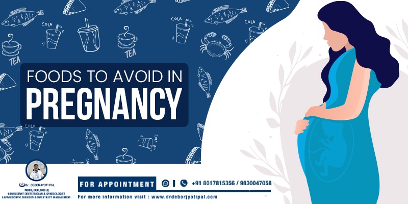
• Mother should food according to her choice in adequate amounts. It should contain adequate amounts of proteins, carbohydrates, fats, vitamins and micronutrients. Can take advice from nutritionists if needed. Breast feeding mothers need higher caloric intake in their food (500 calories extra than non breast feeding women).
• Adequate sleep is needed. So any problems leading to less sleep for the new mother should be dealt with promptly.
• Constipation after delivery should be treated with food having high leafy vegetable content, lots of oral water intake (3 lit/day) and if needed laxatives can be used.
• Mother should try and pass urine as early as possible after a vaginal delivery. If passing urine becomes a problem due to pain in stitches or less bladder tone then an urinary catheter is inserted to evacuate urine. The catheter can be kept inserted into the bladder till bladder tone returns. In case of cesarean delivery same principle to be followed after removing urinary catheter inserted during the operation.
• Post vaginal delivery the vaginal and perineal stitch area should be kept clean & dry. Area should be washed with soap & water after every act of urination & defecation. Wiped with dry clean cloth. For initial 2 weeks an antibiotic ointment can be used to apply on perineal wound.
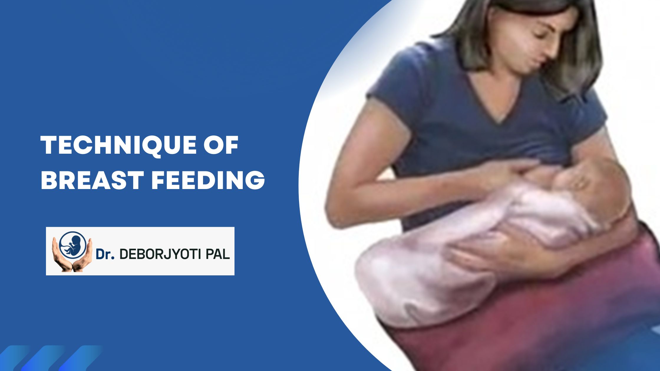
• Breast care

  • Wear a nursing bra that fits well but is not too tight or restrictive, but actually supports the breasts well. Avoid underwired bra.
  • Take daily bath. Wash breast with normal water only. Do not use soap as it causes cracked nipples by removing the protective coating on nipple and areola, derived from natural secretions of glands around the nipples.
  • Nipple should be washed with water and cotton before & after each feed and kept dry between feeds.
  • Change nursing pads if they become soiled or wet.

• Rooming-in

Hospital practice where mothers and normal babies stay together in the same room, all day from after delivery. This is to be practiced as it creates the much needed bonding between the baby and the mother. Helps in early initiation and sustenance of breast feeding. Also helps in making the mother conversant with the babies everyday habits and behaviour helping the mother to better raise their children.

• Vaccines

  • Rubella vaccine if not given before pregnancy & mother in non immune to rubella virus.
  • In Rh (-)ve mothers with Rh(+)ve babies Anti-D immunoglobulin injection has to be given within 72 hours of delivery.
  • TDaP vaccine if missed in pregnancy.

Here I have tried to focus in brief about important aspects of normal post delivery care for mothers. This is certainly not an exhaustive discussion but any other queries can be dealt with adequately by your gynecologist or baby doctor if needed. You can also mail your queries to deborjyotipalqueries@outlook.com.


Blue-White-Simple-Financial-Tips-Blog-Banner-2-1200x675.jpg
04/Oct/2023

Definition: The production & secretion of milk by mammary glands in the breast.

This happens in 4 stages during pregnancy and after delivery.

  •  During pregnancy breast tissue proliferates resulting in increase of ducts and sacs for storage of milk needed for breast feeding. This is the stage of MAMMOGENESIS.
  • Milk secretion occurs into the numerous sacs developed in the previous stage under the influence of a hormone- prolactin, after the delivery of baby. This occurs around 3 to 5 days after delivery. This does not occur in pregnancy as high levels of estrogen & progesterone hormone inhibits the effect of prolactin. After delivery levels of estrogen & progesterone decrease substantially. This is the stage of LACTOGENESIS.
  • This is the stage when milk secreted into the numerous small sacs in breast tissue is pumped out into the bigger ducts of the breast near the nipple from where it can be expressed by mother or the child can suck it. Happens due to contractions of minute muscles in the sac walls under the influence of OXYTOCIN hormone. This is produced in the mothers brain when the baby sucks on the mother’s breast. So frequent suckling of breast by baby is essential for milk ejection from breast. This is the stage of GALACTOKINESIS.
  • Sustenance of milk production is possible only by regular suckling of maternal breast by baby following proper technique. Avoidance of mental stress and breast engorgement is essential for this stage of GALACTOPOIESIS.

IMPORTANT STEPS FOR EARLY INITIATION OF MILK PRODUCTION:

  • Put the baby to mother’s breast within 1 hour of birth.
  • Encourage mother to drink plenty of fluids.
  • Let the baby suck mother’s breast every 2-3 hours as needed.
  • Avoid mental stress, breast engorgement.
  • Completely avoid feeding anything else to the baby (EXCLUSIVE BREAST FEEDING).
  • Avoid bottle feeding.

TECHNIQUE OF BREASTFEEDING:

Support the baby on the arm of the same side as the breast you want to feed from. Sit up straight — preferably in a chair with armrests. Cradle your baby in an arm, with your baby’s head resting comfortably in the crook of your elbow while he or she faces your breast. For extra support, place a pillow on your lap.

For any problems or queries about breast feeding consult your gynecologist or pediatrician for proper management.


WhatsApp-Image-2022-12-07-at-16.22.53-1200x600.jpg
07/Dec/2022

Pregnant Mother With Negative Blood Group

WHAT IS A NEGATIVE BLOOD GROUP?

To understand this simply, you need to know that red blood cells(RBC) in the human body have certain proteins on their surface. There can be many types, but the most predominantly used blood grouping system “ABO blood grouping” uses proteins by the name A & B.

When an RBC has an ‘A’ protein on them is blood group A, with a ‘B’ protein on them is blood group B, with both proteins on them its blood group is ‘AB’ and with neither of these proteins present on them, its blood group is ‘O’.

There is also another protein known as ‘RhD’. If this is present on the RBC then the blood group is ‘+’ve. If this protein is not present then the blood group is negative.

So if you have ‘A’ protein on your RBC and no ‘RhD’ protein, then the blood group is ‘A (-)ve’. Every other blood group is determined this way.

WHY IS IT IMPORTANT?

Now we need to know that if a (-)ve blood group woman marries a (+)ve blood group man and gets pregnant with him, then there is a chance that the baby in the womb may have (+)ve blood group. If that is the case then in the first pregnancy, some RBCs of the baby pass into the mother’s blood due to various reasons (elaborated later), which

produces antibodies against the ‘RhD’ protein on the baby’s RBC.

This antibody development if not prevented can cause dire consequences in future pregnancies, causing severe anemia in the baby and also the death of the baby before or after delivery in all future pregnancies.

SOME REASONS FOR A BABY’S BLOOD MIXING WITH MOTHER’S?

  • Induced
  • Spontaneous miscarriage.
  • Any bleeding inside the uterus during
  • Any diagnostic procedure like Amniocentesis, Chorionic villous biopsy.
  • Normally at certain times in the pregnancy.

GYNECOLOGIST’S ROLE?

Pregnant women with negative blood groups should therefore always be under regular supervision during their pregnancy, giving the gynecologist adequate opportunity to detect the development of these antibodies & also take preventive measures against their development.

To know more about this condition and seek help you can book an appointment with me by calling on 9830047058/8017815356 OR you can go to the appointments section on deborjyotipal.com

Read our other blog What is Poly Cystic Ovarian Disease?-MYTHS & REALITIES


WhatsApp-Image-2022-10-07-at-13.40.02.jpeg
07/Oct/2022

In continuation of my earlier blog in the series ‘USG IN PREGNANCY (PART 1)’, I will give information about the scans done between weeks 13 to 29 of pregnancy, and their clinical importance.

  • 18-20 weeks –

    ANOMALY SCAN’. As the name suggests we try to find out any anomalies or abnormalities in the structural growth of the baby. External and internal organs are checked for anomalies.

  •  22-24 weeks—

‘CERVICAL LENGTH’ Studies have proved that the length of the cervix (the lower opening of the uterus from where the baby comes out in a normal delivery), has a good predictive value for preterm vaginal delivery at around 28-30 weeks. The general consensus is that the length of the cervix should be more than 2.5 cm at this time & should not be showing constant decreasing length from earlier scans for assurance about chances of early delivery being high.

FETAL ECHOCARDIOGRAPHY’ is also done during this time to recheck if the heart of the baby has no anomalies (if it had been missed earlier during the ANOMALY scan).

There is also a way to predict the development of maternal high blood pressure and slow growth of the baby in late pregnancy, by seeing the speed of flow of blood and its variations in the arteries of the uterus at this time of the scan.

  • 28 weeks—

    This usg is done to find out if the baby is growing well corresponding to the age of the pregnancy. Moreover, the amount of fluid around the baby, baby weight, placental maturity, baby movements, breathing actions, and placenta location changes in case of the low-lying placenta in earlier scans are some of the other important things noted.


 In my 3 part blog on this topic, I have elaborated on ultrasonographic in various phases of pregnancy and each of their importance. To know more on this topic you can mail me at deborjyotipalqueries@outlook.com.

Read USG In Pregnancy Part 1 Here

You can also book appointments with me by calling on 9830047058/8017815356 OR you can go to the appointments section on www.deborjyotipal.com.

Follow our Facebook page for more – Dr. Deborjyoti Pal


WhatsApp-Image-2022-10-07-at-11.40.52.jpeg
07/Oct/2022

  • WHAT IS USG?

USG or Ultrasonography is a very commonly used investigation in pregnancy. The basic mechanism is that sound waves are emitted by the usg probe placed on the abdomen or inside the vagina, which gets reflected back to the probe from the various structures of the baby and around it, to then be converted into an image on the usg machine screen.

Many patients fear that there are harmful rays in usg that may harm the fetus. But there are no such rays in usg, I can assure you all of that.

  • PURPOSE: usg done at specific times during the pregnancy period to get information about how the pregnancy is developing, detect the possible presence of developmental defects both at structural and genetic levels & predict the development of certain harmful conditions in the developing baby, so that actions can be taken to prevent those harmful conditions from developing.
  • TIMINGS & PURPOSE OF EACH USG:

  • 6-8 weeks—Known as the ‘DATING SCAN’. It accurately gives the age of the fetus. Moreover, it detects for the first time the presence of a heartbeat in the baby. Importantly it also detects the number of developing babies and the exact location of the pregnancy. (Pregnancy developing in any location other than inside the uterus is known as ECTOPIC pregnancy & is very dangerous for the mother, sometimes leading to death if not detected early enough or not treated properly)


  • 11-14 weeks—‘NT SCAN’. Simply speaking, the length of a translucent area at the back of the baby’s neck is measured. Also, the presence of nasal bone is looked for. Certain blood tests are done with maternal blood. All these data along with the maternal age at conception is taken into consideration for calculating the probability of the presence of certain chromosomal disorders and structural defects in the developing baby. Reported as ‘HIGH RISK’ or ‘LOW RISK’. High-risk mothers are evaluated more for coming to a definitive diagnosis.

Also, the location of the developing placenta (the organ that is responsible for supplying all nutrition and oxygen to the baby) is noted in this scan. Location is important as the placenta develops in the lower part, close to the lower opening of the uterus is a major concern, as it can result in repeated minor/major bleeding episodes in pregnancy, which keeps the gynecologist on his/her toes.

Read USG In Pregnancy Part 2 Here

In my 3 part blog on this topic, I have elaborated on ultrasonography in various phases of pregnancy and each of their importance. To know more on this topic you can mail me at deborjyotipalqueries@outlook.com.

You can also book appointments with me by calling on 9830047058/8017815356 OR you can go to the appointments section on www.deborjyotipal.com.


WhatsApp-Image-2022-09-23-at-18.34.19.jpeg
23/Sep/2022

DEFINITION—

Acute and sudden development of inability to pass urine on one’s own will.
Sometimes it is accompanied by lower abdominal pain due to overdistension of the urinary bladder with huge amounts of stored urine if the inability persists for an appreciable amount of time.
Acute urinary retention occurs in approx. 4 to 5 pregnant women out of every 1000. In some studies, its occurrence is as low as 1 in 3000 pregnancies.

TIME OF OCCURRENCE—

Mostly occurs between the 10th to 16th week of pregnancy.

CAUSES—

• Most commonly there is dysfunction of urethral sphincter relaxation.
• Woman aged 35 or more during pregnancy.
• Previous abortion.
• Endometriosis.
• Recurrent history of pelvic infection.
• Women born with deformities of the uterus.
• Fibroid uterus.
• Abnormal shape of the pelvis.
• Urinary tract infection.

SYMPTOMS OF ACUTE URINARY RETENTION—

• Distress in passing urine with decreased flow requiring more effort.
• Stop starting flow during passing urine.
• Non-passage of urine causing bladder distension and pain in the lower belly.

CONSEQUENCES-

In advanced cases bladder rupture, hydronephrosis, Acute renal failure, and spontaneous abortion.

WHAT TO DO REGARDING ACUTE URINARY RETENTION IN PREGNANCY—

Early recognition of these symptoms and information passage to your gynecologist as early as possible is a must, to avoid severe consequences. Hospitalization and continuous catheter placement inside the bladder might be necessary for 48 hours. If managed properly and on time severe complications can be easily avoided. Post-discharge from the hospital, you might be prescribed certain exercises, which need to be done without fail.

To know more about this and seek help you can book an appointment with me by calling on 9830047058/8017815356 OR you can go to the appointments section on www.deborjyotipal.com.


WhatsApp-Image-2022-09-19-at-11.14.04.jpeg
19/Sep/2022

WHY DO POSTPARTUM EXERCISE?

• Increase strength & tone in your abdominal & pelvic muscles.
• Lose weight.
• Can help to attain good sleep.
• Postpartum Exercise refreshes the mind & keeps mental stress away.

MUSCLE CHANGES IN PREGNANCY

• Anterior abdominal wall muscles move apart through the midline, as the uterus fills up the abdomen throughout the pregnancy. They also lose their strength and tone.
• Pelvic floor muscles and ligaments that support the bladder, uterus and bowel – can be weakened after pregnancy, especially if you had a large baby or had a difficult and prolonged labor process.
Regaining strength and tone in these muscles is essential after pregnancy is over to prevent herniation of internal organs through the gaps in the muscles or less supporting strength of the muscles.

HOW TO DO POSTPARTUM EXERCISE AND FROM WHEN?

Within a week after delivery (Vaginal or Caesarean), abdominal wall and pelvic floor muscle strengthening exercises can be started. Walking can be started in case of vaginal delivery, slowly increasing it to up to 30 min walk per day, totalling 150 mins a week, according to individual comfort. Light walks inside your house is ok after caesarean delivery. Outdoor walking needs to wait till up to 6-8 weeks of caesarean.

Abdominal wall muscle exercise—

1. Lie on your back with slightly bent knees
2. Gently pull in your lower abdomen and your pelvic floor while exhaling.
3. Hold in for 10 secs and gently release them.
4. Repeat points 2 & 3 ten times.
5. 4 times a day.
Pelvic floor exercise—
1. Sit and lean slightly forwards.
2. Squeeze in the muscles around your vagina, as if trying to control the passage of stool or urine.
3. Hold for 10 secs and gently release.
4. Repeat points 2 & 3 after 10 secs relaxation, 10 times in total.
5. 4 times a day.

For better information about these contact your physiotherapist or gynaecologist.

After 6-8 weeks of delivery (vaginal or caesarean), you can start going to the gym, but only for certain light exercises—

• Low impact aerobics.
• Cycling.
• Yoga.
• 30 min/day outdoor walks (in cases of caesarean delivery).
• Pilates.
• Light weight training.
• Swimming after 7 days of stoppage of per vaginal bleeding.

After 4 months of delivery (vaginal or caesarean)—

• High-intensity aerobics.
• Running.
• Heavyweights & sit-ups.

POSTPARTUM EXERCISE FOR BREASTFEEDING MOTHERS –

• Do not exercise with full breasts. It can be very uncomfortable. Breastfeed or pump out milk beforehand.
• Do not do very strenuous exercise. Breast milk quantity might decrease.
• Wear a well-supportive bra. Neither too tight nor too loose.
• Drink plenty of water.

WHENEVER EXERCISING, KEEP IN MIND TO STOP DOING ANY ACTIVITY IF IT CAUSES DISCOMFORT OR PAIN & TO STOP THAT SPECIFIC ACTIVITY FOR A FEW DAYS & START SLOWLY LATER.

To know more about this and seek help you can book an appointment with me by calling on 9830047058/8017815356 OR you can go to the appointments section on www.deborjyotipal.com.

Visit Our Facebook Page Here Dr. Deborjyoti Pal


WhatsApp-Image-2022-09-16-at-12.52.32.webp
16/Sep/2022


nausea and vomiting during pregnancy is a commonly encountered symptom typically starting around 5th to 7th week and resolving by the 5th month of pregnancy in 90% patients. It’s seen in (50 – 80) % of pregnant women.

A special clinical form of this is HYPEREMESIS GRAVIDARUM. Characterized by continual symptoms even on medication causing dehydration and significant weight loss in patients.

Sisters and daughters of patients with this condition have a higher chance of developing this condition in pregnancy.


Certain health conditions also have the same symptoms of –

  • Diabetic complications, Thyroid disease.
  • Migraine, Brain tumors, Ear diseases.
  • Hepatitis, Appendicitis, Pancreatic problems, Excessive acidity.
  • Psychiatric disorders, Drug side effects.
  • High BP in pregnancy, Liver disease in pregnancy.
  • Ovarian cyst complications, Uterine fibroid complications.
  • Urinary tract infection, Kidney stone.


Some patients are more at risk of nausea and vomiting during pregnancy—

  • Family history.
  • Previous personal history in earlier pregnancy.
  • Pregnancy with more than one fetus.
  • Abnormal pregnancy like Molar pregnancy.
  • History of migraine, motion sickness.
  • Low BMI.
  • Female baby.


Non-pharmacological methods to get some relief from nausea and vomiting during pregnancy—

  • Lifestyle changes –

  1. Small & frequent meals (6 meals a day)
  2. Meals should be high in carbohydrates & protein and low in fat content.
  3. Bland diet.
  4. Avoid keeping empty stomach. Eat whenever hungry.
  5. Avoid smells or foods that cause vomiting.
  6. Avoid stuffy rooms, hot places, noisy surroundings, driving on rough & undulating roads, excessive exercise.
  7. Avoid lying down immediately after eating.
  8. Drink adequate amounts of water. 2.5 lits/day should be the aim.


  • Ginger containing foods-

Small amounts of cold, clear ginger ale in between meals might help. Ginger lollipops can be useful


  • ACCUPRESSURE can be helpful in some.

WHEN SHOULD YOU SEEK MEDICAL HELP—

  • Incurable vomiting not responding to oral medications.
  • Loss of weight (2-3) kgs.
  • Dark colored urine.
  • Abdominal cramps.
  • Depressed mental state/ Thoughts of Terminating pregnancy/ Suicidal thoughts.


To know more about nausea and vomiting during pregnancy and seek help you can book an appointment with me by calling on 9830047058/8017815356 OR you can go to the appointments section on www.deborjyotipal.com

Follow Our Facebook Page: Dr. Deborjyoti Pal


WhatsApp-Image-2022-08-27-at-10.57.59-AM.jpeg
27/Aug/2022

When you’re pregnant, you learn about all the foods that are off-limits. It can be hard if you love sushi, coffee, or steak. Thankfully, there are more things you can eat than things you have to avoid. To stay healthy and safe during pregnancy, here are 6 foods  to avoid or limit .

1. Avoid Seafood high in mercury during pregnancy 

Some fishes like tuna, Swordfish, king mackerel, shellfish, shark contain high levels of mercury. Avoid bigger and older fishes.Upto 350 gms of sea food a week can be allowed.Catfish, trout, tilapia, salmon, shrimp, cod, herring are
some of the fishes allowed.

Avoid raw, undercooked or contaminated sea Food. Avoid refrigerated sea food . Fishes to be cooked
Preferably to 65 degrees celsius. Fishes to be cooked till Its opaque . Oysters or mussels to be cooked till their
Shells open.

2. Herbal teas to be avoided during pregnancy

There is very little safety data about herbal tea effects on developing babies. So avoidance is safe
.

3. Caffeine Intake During Pregnancy 

Upto 200mg can be allowed a day. For perspective a cup of coffee(250ml) has 95mg same amount of tea has 50mg and 350ml cola has 30mg caffeine.

4. ALCOHOL is off -limit

Not safe in any amount in pregnancy. Avoid completely.

5. What cheeses to avoid during pregnant?

Soft cheese like feta, brie, blue cheese should be avoided. Unpausterized milk should also be avoided.

6. Avoid Undercooked meat, eggs, poultry while pregnant

Undercooked meat can cause fatal listeriosis infection .  Eggs should be completely boiled with hard yolk and egg white. Hollandaise sauce, Caesar salad dressing may contain raw egg components and should be avoided.

7. UNWASHED fruits & vegetables and RAW SPROUTS 

To know more about food habits and any other queries you may have, you can post your queries in the QUERIES section on www.deborjyotipal.com or can fix an appointment by calling on 8017815356 for face to face interaction. You can also follow Dr. Deborjyoti Pal on Facebook to get the latest gynecology updates news and tips.


WhatsApp-Image-2021-06-24-at-12.38.04-1.jpeg
25/Jun/2021

There can be a number of reasons because of which a miscarriage can occur. Repeated pregnancy loss or recurrent miscarriage can be defined as two or more consecutive pregnancy losses before the pregnancy reaches 20 weeks of age. By finding out the root cause behind the miscarriages and following the right path of treatment can often solve these difficulties efficiently.

It is extremely vital to know that the majority of women require therapy and the right treatment for dealing with these odds and for getting a legitimate solution for diminishing the chances of pregnancy loss. Dr Deborjyoti Pal is one of the best gynecologist in Kolkata who is well-known for treating complicated issues with recurrent pregnancy loss.

Here are some of the possible reasons because of which women tend to have repeated miscarriage:

● A structural problem with the uterus

An ultrasound or an X-ray test can find out the structural abnormalities of the uterus in no time. It can be polyps, fibroids, congenital malformations etc. that are leading to complexities in the pregnancy. In most cases, these conditions can be dealt with surgery. Therefore, there is no need to worry as one can opt for affordable infertility treatment in North Kolkata without any difficulty.

● Hormonal disorders

Thyroid disease, high prolactin levels or uncontrolled diabetes can often increase a woman’s risk of pregnancy loss. Therefore, it is highly recommended to treat these problems first and then to proceed ahead to having a safe journey with pregnancy.

● Blood clotting problems

Certain substances by there excessive pesence or absence in blood can lead to formation of small clots in the placenta of the baby causing blockage of small blood vessels in the placenta and stoppage of supply of nutrition and oxygen to developing baby in the womb, thus causing early miscarriage or late intrauterine death.

● Infections

Certain viral, parasitic, bacterial organisms can lead to this condition although this is a rare/doubtful causation.

● Chromosomal

Certain chromosomal abnormalities in the parents or the developing baby can cause this condition.

Conclusion

Besides these, there are a number of other complications which can not be detected at the first chance with ease. That is why it is extremely necessary to consult a medical expert for finding a valid solution. Dr Deborjyoti Pal is a gynecologist near Sodepur who has been dealing with patients suffering from repeated miscarriages for years. One can definitely consult him and seek advice for sorting out the complications effectively.

Follow our facebook page: www.facebook.com/drdeborjyotipal


Dr Deborjyoti Pal - top gynecologist in Sodepur, Kolkata

Consultant Obstetrician & Gynaecologist


Consultant Obstetrician & Gynaecologist


About Dr. Deborjyoti Pal

MBBS, DGO, DNB(I)

Consultant Obstetrician & Gynaecologist

Laparoscopic Surgeon & Infertility Management

Attached to Bellevue Clinic & Birth Fertility Clinic

+91 98300 47058


24/7 EMERGENCY NUMBER

Call us now if you are in a medical emergency need, we will reply swiftly and provide you with a medical aid.


deborjyotipal@outlook.com


Copyright by Dr. Deborjyoti Pal 2020. All rights reserved. Design by Seven Boats


Copyright by Dr. Deborjyoti Pal 2020. All rights reserved. Design by Seven Boats